Temporomandibular joint study
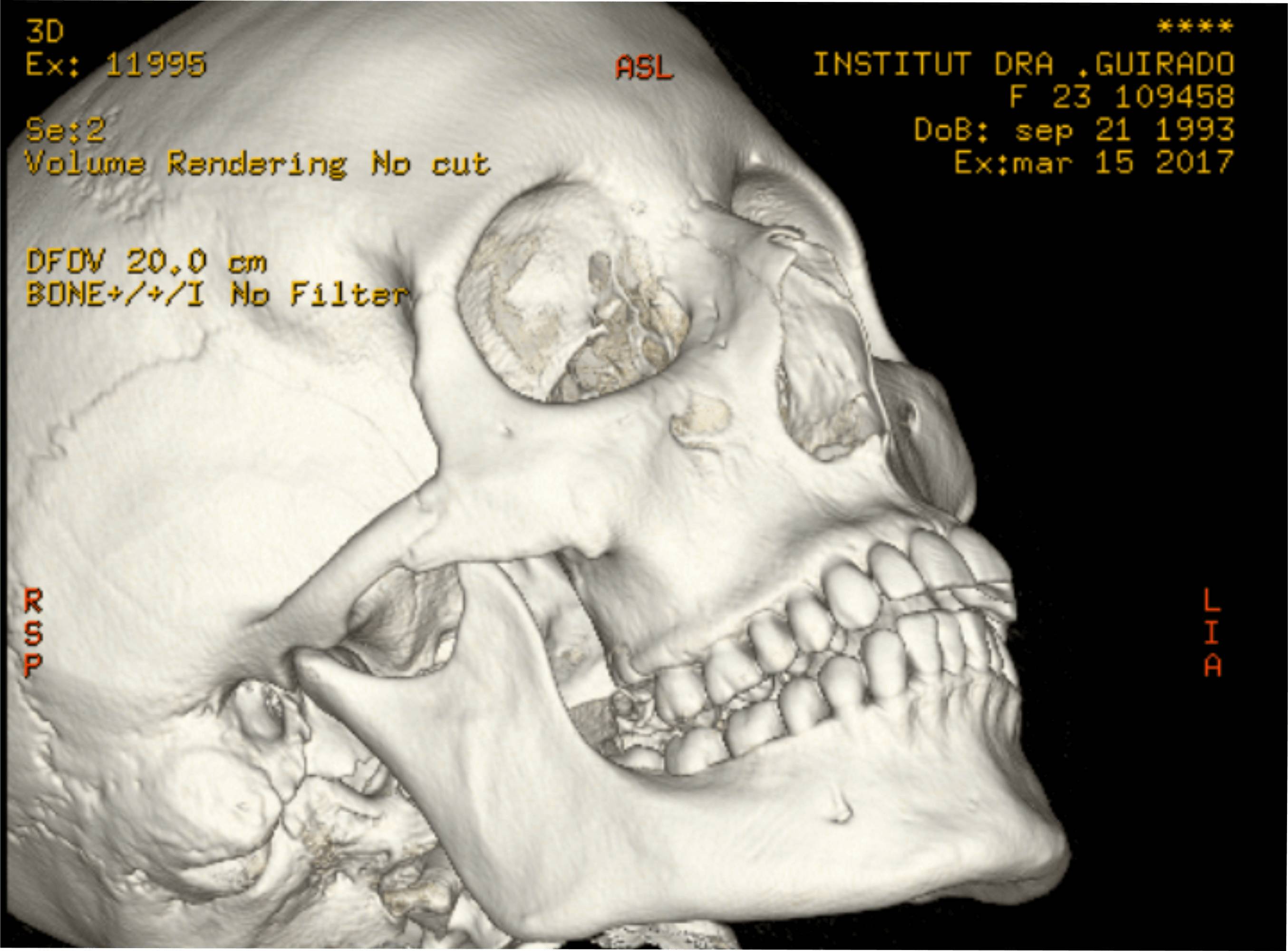
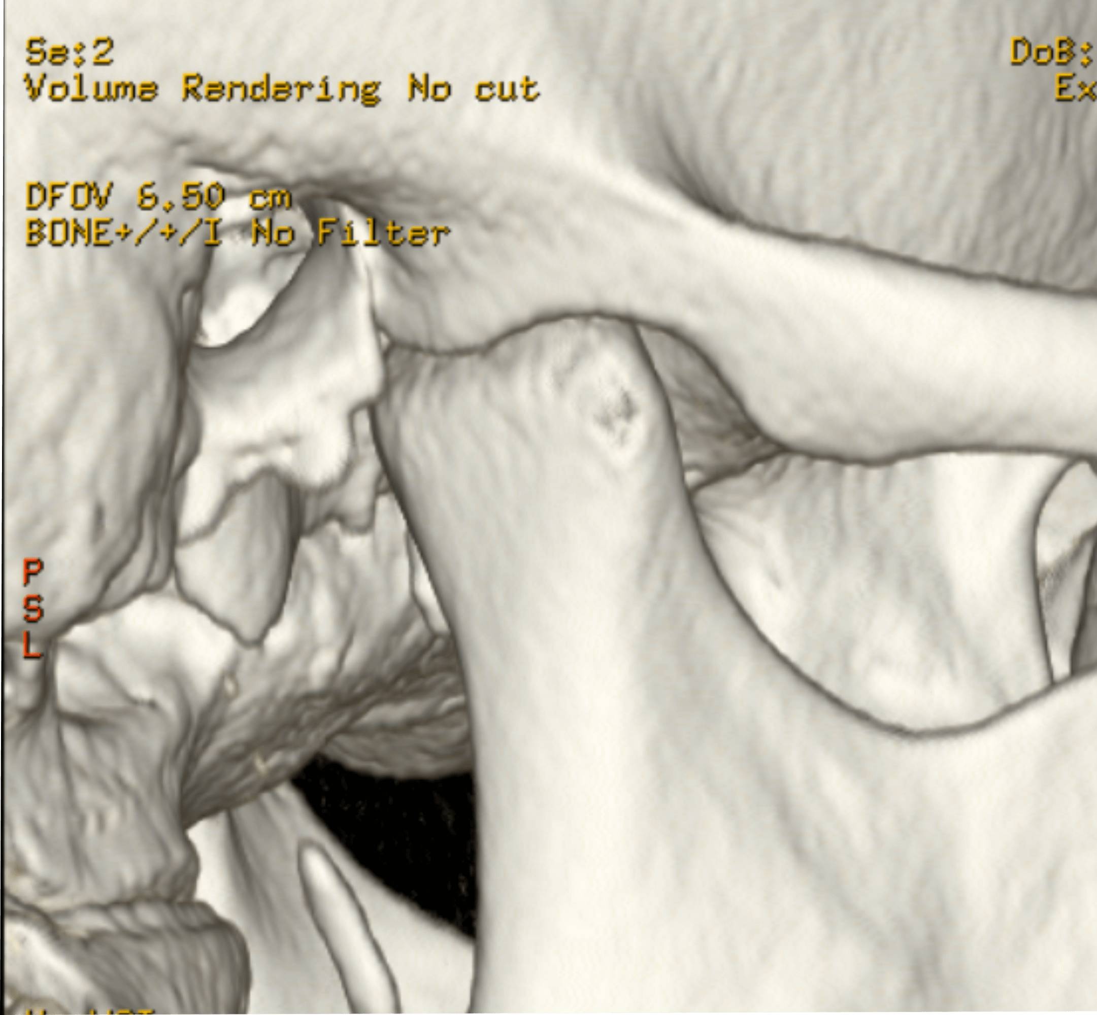
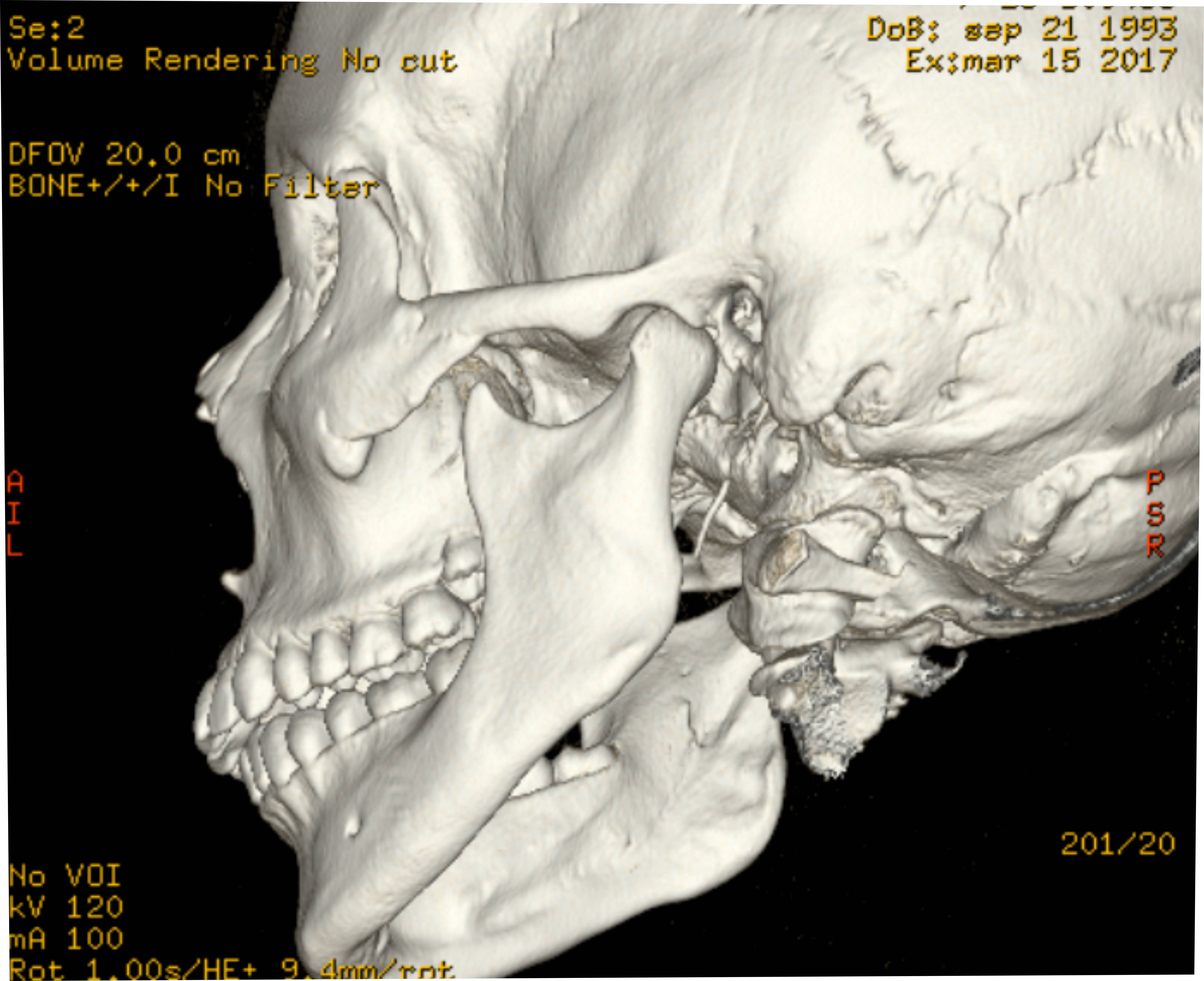
< Go back to "Diagnostic from Dental Imaging"NewTom 5G XL improves the diagnosis of M.T.A. Sagittal and coronal slices provide an excellent representation of the joint space and allow to detect the presence of pathologies. The 3D rendered images show a high quality and accuracy, thus favoring the anatomical evaluation of the A.T.M. and allowing other important evaluations, such as the difference between the height of the condyle and the mandibular ramus, to be carried out. Using contrast medium, the meniscus can also be examined.
Examples of cases